- 2026/09/20
- مقالات خارجی
- نظرات
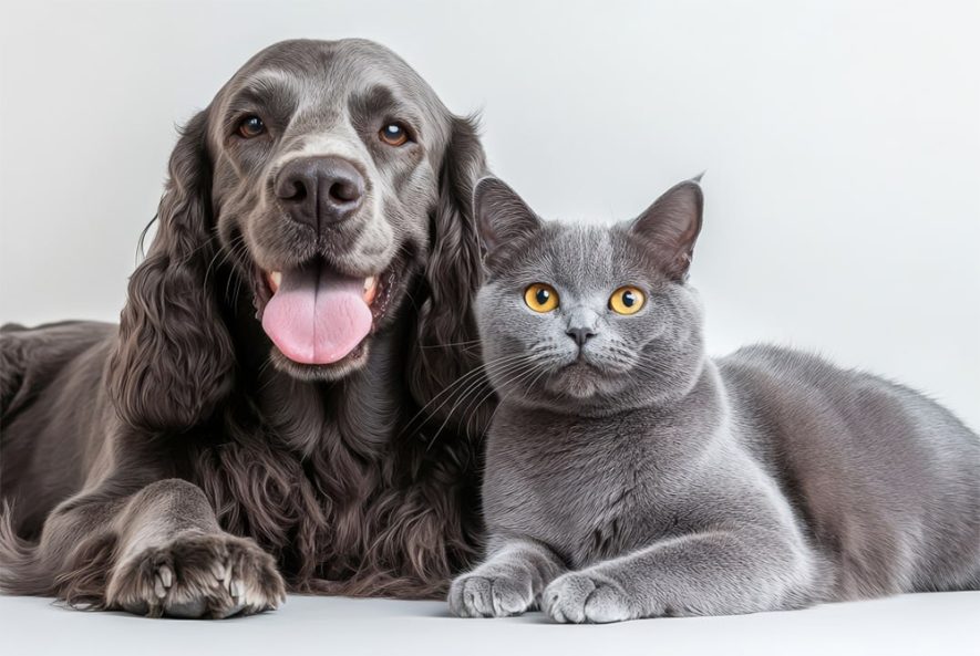
The skin is the largest organ of the body with many different functions as thermoregulation, immune protection, sensory perception, vitamin D production and it acts as a barrier between the animal and the environment. Besides all of these important functions and the diseases that affect directly the skin, it may also share or reflect pathologic processes from other tissues. Due to these characteristics, dermatologic problems are among the most commonly seen disorders in veterinary hospitals. It is important for the veterinarian to know and understand about the physiology of the skin and about the most common dermatologic disorders that affects dogs and cats, which will be addressed in this chapter.
Skin structure
The skin is divided in three layers: epidermis, dermis and hypodermis (Figure 1). Epidermis, the outermost layer of the skin, is composed by keratinocytes, melanocytes and Langerhans cells. Keratinocytes are also disposed in layers in the epidermis. The deepest one, the stratum basale, is formed by a single row of germinative keratinocytes and also contains melanocytes. These germinative keratinocytes generate the other layers by cell division and differentiation. The next layer, stratum spinosum, differs from stratum basale by the presence of intercellular junctions. Langerhans cells are also present in this layer. Stratum granulosum is characterized by a large amount of keratohyaline granules inside keratinocytes which are important in the skin keratinization process. Stratum corneum, the outer epidermis layer, is composed by keratinocytes in their maximal differentiation degree (corneocytes), interspersed in lipid matrix [1].
Dermis, the layer under the epidermis, is composed by a conjunctive matrix where reticular, elastic and collagen fibers are found. Dermis cellular structure is composed by fibroblasts, mast cells and histiocytes. It also contains epidermal appendages (hair, nails, sebaceous and sweat glands), arrector pili muscles and blood and lymph vessels. Hypodermis or subcutaneous tissue provides support and cushioning against physical trauma. It is composed by a loose connective tissue and elastic fibers interspersed by adipocytes [1]. The hair follicles exhibit activity cycles that result in hair formation. Anagen is a period of active growth when a new hair is being formed. Catagen is when the hair growth stops and degenerative changes occur in the base of the follicle. Telogen represents a period of follicle inactivity, when the hair is shed so that a new one may start to grow [1]. Hair cycle activity, in some dog breeds, is strongly related to temperature variation and photoperiod, leading to decreased hair density in the warmer months, which helps the heat loss in these animals [2].
Diagnosis techniques
Diagnosis approach for skin diseases depends on obtaining detailed history with thorough physical and dermatological examination. The evaluation offers precious information and guides the investigative process. Specific tests and, occasionally, therapeutic trials should be performed [3].
Skin scrapings, trichogram, fungal and bacterial culture, cytological evaluation and skin biopsy are important diagnosis techniques in dermatology. Skin scrapings are applied in mite detection, and it may be superficial or deep depending on the mite that is suspected. The sample obtained is evaluated under optical microscopy [3]. Fungal culture is recommended for patients that fungal diseases are suspected. Hair samples and skin scales should be collected from the lesion margin. Bacterial culture is not often performed and it is recommended in pyoderma refractory to initial therapy or when rods are observed in cytology [4]. The direct examination of the hairs, the trichogram, is performed by pulling the hairs from the affected area, followed by microscopic evaluation. With this technique it is possible to determinate hair growth phase abnormalities and the presence of follicles parasites and dermatophytes [3]. Cytology is the analysis of tissue cells and it is a highly efficient and valuable exam to evaluate a lesion, after which it is possible to establish the next step in the diagnostic approach. The sample may be obtained by fine-needle aspiration, swab, skin scrapings or lesion imprint [5]. Histopathology associated with clinical findings usually leads to definitive diagnosis. Skin biopsy is recommended in unusual lesions, possibly neoplastic nodules, dermatosis with expensive therapy or when it represents a risk to the patient health, with poor response to previous therapy and to exclude differential diagnoses [4]. To perform the histopathological exam at least three representative samples should be obtained by punch or surgical resection [3].
Bacterial skin diseases
Pyoderma is a bacterial skin infection and it is among the most common causes of skin diseases in dogs [6], however, it is less common in cats [7]. Lesions may be superficial and involve only the epidermis or they may affect deeper structures in the dermis or subcutaneous tissue, and it is therefore divided into surface, superficial and deep pyoderma [6]. Staphylococcus pseudintermedius is the most commonly isolated bacteria from dog’s skin [8] and it is among the main reasons for antimicrobial use in these animals [9].
Surface pyodermas are characterized by superficial erosions of the stratum corneum. The presence of alopecia, erythema and pruritus are common findings [10]. Intertrigo is a surface pyoderma that affects the skin folds found in lips, face, vulva, tail and mammary glands of some breeds, and it may also affect the skin folds of obese animals. Acute moist dermatitis, also known as pyotraumatic dermatitis or hot spots, is of acute onset and rarely occurs as a primary disease in healthy skin, being usually secondary to other diseases [11]. Probably, local irritation due to an underlying cause leads to self-inflicted trauma, which quickly becomes extensive areas of skin damage [10].
Superficial pyodermas are the most common causes of cutaneous bacterial infection in dogs [6]. They affect the superficial portion of the hair follicles (bacterial folliculitis) or the epidermis (impetigo), causing pustules [10, 11]. The most common lesions are crusted papules due to the transient nature of canine pustules. Pruritus, epidermal collarettes, hyperpigmentation and alopecia are also common findings [6]. Impetigo affects sexually immature dogs that may present subcorneal pustules formed in inguinal and axillary areas [10]. Superficial bacterial folliculitis is the most common form of pyoderma in dogs [12]. Papules, pustules associated with hair follicles, epidermal collarettes, alopecia and hyperpigmentation are commonly found [10] (Figure 2).
Deep pyoderma does not occur spontaneously, often starting as superficial pyoderma [6]. Other organisms such as Proteus spp., Pseudomonas spp. and E. coli may be involved [12-14]. The bacterial infection affects the deepest portion of the hair follicle (deep folliculitis), that may lead to follicular wall rupture and to bacterial product release in the dermis (furunculosis), or it can also affect the deeper portion of the dermis and subcutaneous tissue (cellulitis) [10, 15]. The affected skin appears erythematous, hyperpigmented, with the presence of seropurulent debris from the ruptured pustules; variable pruritus, swelling, skin stiffness and evident pain are also noted [6].
Diagnosis is obtained through the evaluation of clinical signs, presence of characteristic skin lesions, elimination of other possible causes of folliculitis and by cytological evaluation of the intact pustules content, exudative lesions and skin debris. In the management of pyoderma, it is important to identify the possible underlying disorder, which may be done through skin scrapings, scabies therapeutic trial, allergy tests, endocrinopathies screenings, hypoallergenic diet trials, strict ectoparasites control and skin biopsies. [16].
The treatment varies depending on presented lesions. Local surface and superficial pyodermas may be treated only with topical antibiotics such as silver sulphadiazine, neomycin or 2% mupirocin ointments applied twice daily over the affected areas. Generalized lesions and deep pyodermas require a combination of oral and topical antibiotics. In patients with severe pruritus it is recommended to use anti-inflammatory doses of prednisone orally for up to two weeks [17]. The antibiotics of choice for oral use include cephalexin (22-33 mg / kg q12h) and amoxicillin associated with clavulanic acid (22 mg/kg q12h) [18]. Recurrent cases require culture and susceptibility testing to access resistance [16].
Fungal diseases
– Dermatophytosis
Dermatophytosis is a superficial mycosis caused by Microsporum, Trichophyton or Epidermophyton fungi genera. These fungi are isolated from hair, nails and skin surface since they require keratin for their growth [19]. Dermatophytes are classified into three groups based on their habitat: zoophilic, mostly found in animals, occurring transmission to other animals or to humans; anthropophilic, mostly found in humans, transmitted between humans and rarely to animals and geophilic, dermatophytes, found in the soil, infects humans and animals [20]. M. canis is the most frequently isolated fungal species in dogs and cats [21].
Clinically, canine and feline fungal infections differ. Infections in dogs often produce lesions, while it is possible to isolate dermatophytes from clinically healthy cats, which can act as a conidia reservoir of the fungus [22]. The affected animals usually have alopecic, scaly, crusted, erythematous and papular lesions, especially in the face and limbs. Occasionally, dermatophytes may be presented in a nodular form known as kerions. This form of dermatophytosis is characterized by deep, inflammatory and suppurative lesions [23]. Pruritus may vary from absent to severe [24].
The direct microscopic examination of hairs and scales can reveal the presence of fungal hyphae or spores. The fungal culture is the diagnostic test of choice and the sample may be obtained by brushing the animal with a toothbrush or by skin scrapings [25].
The best strategy for the treatment of dermatophytes is the association of systemic and topical antifungal therapy. The aims of the treatment with topical products are the elimination of the fungi present at the epidermis and hair surface, while systemic treatment aims to eliminate infection within the hair shafts [26]. Lime sulfur rinse at 6.5 % twice a week showed good results in cats infected with M. canis [27, 28]. Systemic treatment options include itraconazole orally at 10 mg/kg once a day, griseofulvin 50 mg/kg once a day or terbinafine 5 mg/kg once a day [27-29]. The treatment must be extended over 2 to 4 weeks after clinical cure and after obtaining two or more negative fungal cultures [23].
– Malassezia dermatitis
Malassezia pachydermatis is a commensal skin yeast, commonly isolated from lips, interdigital skin, anal mucosa and external auditory canal [30]. It is an opportunistic yeast, which usually manifests itself after the installation of other diseases. It is very common in dogs and least frequent in cats [31]. Previous antibiotic therapy is associated with the development of cutaneous M. pachydermatis over growth in dogs [32], as well as disorders of keratinization and hypersensitivity diseases [33]. Basset Hounds, Cocker Spaniels and West Highland White Terriers are more predisposed to this type of infection [32].
The most common clinical manifestation of Malassezia dermatitis is the presence of moderate to intense pruritus [34]. Erythema, lichenification, oily skin, malodor, alopecia and erosions are also common clinical findings [35] that can be generalized or localized [34].
Cytology is the diagnostic method of choice and allows microscopic identification of the increased number of yeasts. Samples can be obtained by skin scrapings, swabs, direct imprint or by tape preps. Fungal culture is not recommended as a diagnostic procedure because it is not a quantitative assessment [36].
Identification and correction of the predisposing diseases are essential for the successful treatment of Malassezia dermatitis [23]. Mild cases can be treated with miconazole, chlorhexidine, ketoconazole or acetic acid shampoos. Baths should be given, two times per week, for a minimum of six weeks [36]. Patients requiring systemic treatment should receive oral antifungals such as ketoconazole, itraconazole or fluconazole 5 mg/kg once a day for a minimum of 30 days [36]. Terbinafine 30 mg /kg every 24 hours on two consecutive days in a week, for six weeks, may also be an effective treatment [37]. In order to prevent recurrences of the disease, regular maintenance therapy may be needed in many dogs [23].
– Sporotrichosis
Sporotrichosis is a subcutaneous mycosis caused by a dimorphic fungus, Sporothrix schenckii, which can infect animals and humans [39]. It is a zoonotic disease and transmission to humans occurs through bites or scratches, and contact with cats ulcers [40]. S. schenckii is present in the decaying vegetation and soil and animal contamination occurs by skin open lesions such as perforations, bites and scratches. Once in the host organism, the fungus may cause local lesions and possibly systemic signs [41].
Clinically, sporotrichosis has three forms: cutaneous, lymphocutaneous and generalized, and more than one form can occur simultaneously in the same animal. The cutaneous form is usually confined to the area of fungus inoculation and manifests after an incubation period of one month. If this lesion is not treated, the progression to lymphocutaneous form can occur. The lymphocutaneous form is characterized by the development of nodules that evolve into ulcers, affecting skin, subcutaneous tissue, lymph vessels and regional lymph nodes [41]. History of lethargy, anorexia, depression and fever on physical examination suggest the presence of the disseminated form [42].
The diagnosis is based on clinical history, physical examination, cytological evaluation, fungal culture and histopathological findings [43]. Cytological evaluation usually reveals oval to elongate yeast cells consistent with S. schenckii form and inflammatory cells may also be present [45]. On histopathology, the presence of deep pyogranulomatous dermatitis, cellular infiltration of polymorphonuclear and mononuclear cells, and the presence of PAS positive structures compatible with S. schenckii may be observed [44]. Oral administration of itraconazole 10 mg/kg every 24 hours is the treatment of choice [46].
– Cryptococcosis
Cryptococcus spp. is a saprophytic fungus present in the environment and in the feces of pigeons, capable of causing systemic infection in dogs and cats, with a higher incidence in felines. The species of interest in veterinary medicine are C. neoformans, which has a global distribution and C. gattii that has a limited distribution [47]. C. neoformans typically infects animals by inhalation and may cause ophthalmic, upper respiratory tract and central nervous system lesions. Ulcerative lesions in the nasal, oral or pharyngeal mucosae, or a nasal masse may be present (Figure 3) [31]. Mycotic rhinitis and cutaneous nasal bridge and nasal plan involvement are the most frequent findings. In cats with positive serology for feline immunodeficiency virus (FIV) cryptococcosis tends to manifest itself in a disseminated or advanced form [48].
Cryptococcosis diagnosis is based on fungal culture, cytological, histological and serological exams [49]. Cytological examination may reveal the presence of leukocytes, macrophages and numerous encapsulated structures (yeast) of different sizes (Figure 3) [50, 51].
Drug therapy leads to patient healing in most cases [49] and it consists of oral antifungal use until complete remission of clinical signs, usually in 3 to 12 months. It is strongly recommended not to interrupt the treatment until the titers of antibodies against cryptococcosis are reduced to zero [52]. Drugs commonly used include fluconazole 50 mg/cat every 12 hours [48] and itraconazole 50-100 mg/cat every 24 hours [52], or 10mg/kg for dogs and cats every 24 hours [49]. Patients with nasopharyngeal masses benefit from surgical resection, for upper airway patency and reduction of infected tissue to be treated medically [52].
Continued article:
https://www.intechopen.com/chapters/43169