- 2026/05/12
- مقالات خارجی
- نظرات

How to Diagnose and Treat It Successfully
Demodex canis is an obligate parasite of hair follicles. A long-body form Demodex injiai and a short-body form Demodex cornei, have also been described in dogs. Cats have two recognized species of Demodex mites, the long-shaped Demodex cati and the short-bodied Demodex gatoi. The latter species is considered potentially contagious to other cats.
Puppies and kitten acquire the infection during the first 72 hours of life, probably while suckling. In dogs there is a definite breed and genetic predisposition to the development of demodicosis; thus, in the case of generalized demodicosis, breeding from affected dogs, their siblings or their parents is discouraged.
Juvenile onset demodicosis (< 1 year of age) is a frequent benign condition in dogs, which is often self-limiting. Adult onset canine and feline demodicosis may occur as a result of immunosuppression, due to glucocorticoid, progestagen or immunosuppressive drug therapy or to systemic diseases, such as spontaneous hyperadrenocorticism, hypothyroidism, systemic infections, neoplasia, malnutrition, parasitism and debilitating systemic diseases. However many of cases of adult onset canine demodicosis have no detectable underlying disease.
Clinical Presentation
Two forms of demodicosis are recognized in dogs:
1. Localized demodicosis is normally seen in animals less than one year of age. The lesions consist of one or two foci of alopecia, commonly on the face or a limb. Some of these cases resolve spontaneously and some may progress to become generalized.
2. Generalized demodicosis can develop from the localized or may appear suddenly. It is characterized by multiple and confluent areas of infestation which may include the feet (pododemodicosis) and the external ear canal (otodemodicosis).
In the absence of secondary infection, and depending on the severity, demodicosis can appear like non-inflammatory patchy to diffuse alopecia, variable erythema and underlying edema, hyperpigmentation, scale and comedone formation. In cases of secondary bacterial infection, lesions observed include pustules, furunculosis, crusts and hemorrhagic bullae. Scarring can lead to permanent hair loss. Pododemodicosis in the milder form presents as interdigital erythema and edema, but then extends swelling, with granuloma and fistula formation. In this form it can be difficult to eradicate, as extensive inflammation and scarring may result in false negative skin scrapings.
Otodemodicosis is nearly always found together with other signs of generalized demodicosis. The otic signs include a ceruminous brown colored exudate with mites on cytological examination.
Pruritus is a variable sign. Cases without secondary infection tend not to be pruritic while those cases with severe secondary infection may display intense pruritus. Systemic signs, such as lymphadenopathy, fever and depression, can accompany severe secondary infection.
Infestation with D. injiai is characterized by greasy skin patches on the dorsum, without hair loss.
Clinical signs of D. cati infection are generally similar to those of canine demodicosis. Alopecia, erythema, crusting, and ceruminous otic discharge can all occur with D. cati infection. Clinical demodicosis due to D. cati infection in cats is often secondary to an underlying systemic disease or immunosuppression. Cats with demodicosis due to D. gatoi infection present with moderate to severe pruritus, self-induced alopecia and scaling on the dorsum, and can be contagious to other cats.
Diagnosis
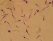
Canine demodicosis is diagnosed by a deep skin scraping performed with either a scalpel blade (size 10 or 20) or a Volkman spoon (5–6 mm in diameter) moistened with a drop of mineral oil. Because demodicosis has a wide spectrum of presentations, examination of a deep skin scraping in all clinical cases is a good practice, even if the diagnosis of demodicosis appears unlikely. Trichograms, performed on hairs collected from the lesions, may detect mites in the keratin infundibular sheaths. After the hair is plucked, it is placed on a drop of mineral oil, covered with a cover slip and examined using 40–100x magnification. In the majority of cases, the number of mites on the host is high and the diagnosis is obvious. In active infections adults, nymphs, larvae and eggs are visible. In cases of otodemodicosis, numerous mites can be seen by examination of a smear of the ceruminous discharge mixed with a drop of mineral oil. Pustules should be examined by cytology for bacteria. The pustule is pricked with a needle and the pus collected as an impression smear. During staining of slides, mites may be lost. Unstained mites, with characteristic body shape, may be seen in the carpet of neutrophils. Bacteria, especially if intracellular, should be noted. If rod-like bacteria are identified on cytology of a pustule then culture and sensitivity testing is indicated, for a correct antibiotic choice.
Occasionally a skin biopsy is indicated in chronic fibrotic lesions, such as the interdigital granulomas associated with pododemodicosis, where a skin scarping is difficult or impossible to perform.
Superficial skin scrapings can be used to detect the superficial mite D. gatoi. Negative scrapings do not definitively rule out D. gatoi infection, so that sometimes empirical therapy for D. gatoi infection is administered to pruritic cats to rule out demodicosis.
Adult onset cases (> 2 years of age) and cats should be rigorously screened for an underlying disease processes by a complete physical examination, hematology, biochemistry, endocrine function tests and tests for internal parasites and for viruses in cats …
Reference:
https://www.vin.com/apputil/content/defaultadv1.aspx?id=5124336&pid=11343#:~:text=Adult onset canine and feline,malnutrition%2C parasitism and debilitating systemic